echnique: LiverApproach the examination of the liver from the right side of the patient. Have the patient lying supine. Preserve the patient’s privacy by draping the top of their body with the gown and below the waist with a sheet. For the best exam, make sure the patient is warm and comfortable. Additionally make sure your hands are warm so as to not startle the patient. InspectionLook for gross asymmetries across the abdomen. Look at the skin for signs of liver disease, such as caput medusa, or spider angiomata. AuscultationFollow the inspection of the liver, as with the rest of the abdominal exam, with auscultation. Listen over the area of the liver for bruits or venous hums. PercussionPercuss for the upper and lower margins of the liver. Place your non-dominant hand palm down flat on the abdomen with the fingers parallel to the lower costal margin pointed toward the midline. Percuss with the middle finger of your dominant hand on the middle finger of your non-dominant one. Begin percussion over the lungs and move from the area of resonant lung sounds to the areas of dullness. Mark the area of change. Repeat the same process from below, moving again from resonance over the bowel to dullness and again mark the area of change. Start in the lower right quadrant so as to not miss a greatly enlarged liver. Measure the vertical distance from the top to the bottom. You can also use palpation to determine the lower border. PalpationBegin palpation over the right lower quadrant, near the anterior iliac spine. Palpate for the liver with one or two hands palm down moving upward 2-3 cm at a time towards the lower costal margin. Have the patient take a deep breath. The liver will move downward due to the downward movement of the diaphragm. Feel for the liver to hit the caudal aspect of your palpating hand. Palpate the bottom margin of the liver for the texture of the liver, i.e. soft/ firm/hard/nodular. Scratch TestSeveral different techniques have been described for this exam. One is to place the diaphragm over the area of the liver and then scratch parallel to the costal margin until the sound intensity drops off marking the edge of the liver. Other techniques involve different patterns of the scratching, for example as in spokes of a wheel and other places for placing the stethoscope such as over the abdomen. Technique: AscitesThere are several physical examination maneuvers described for detection of ascites described below that are at least moderately sensitive and specific. No single maneuver is both highly sensitive and specific; therefore at least two maneuvers are necessary to increase the accuracy of physical exam for ascites. Bulging Flanks
Flank Dullness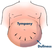
Shifting Dullness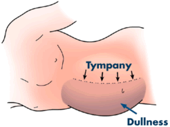
Fluid Wave
| |
feed twitter facebook


9.1.08
Liver
Subscribe to:
Post Comments (Atom)
No comments:
Post a Comment